 VisualNeuroscience.AI
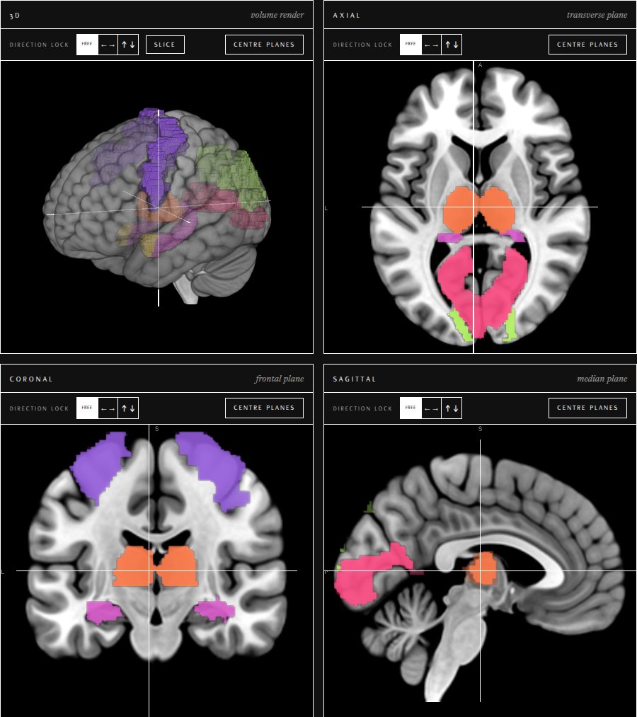
Region Atlas
Pick any of the 116 AAL regions and watch it light up on the MNI152 template at once in axial, coronal, sagittal and 3D. The 41 Brodmann areas live here too: search them by number, by the gyrus they sit on, or by what you want an area to do, pick them off the classic two-face map, and they light up on the same scan in their own colours. Cut the volume open along any axis to see the scan inside the slice, probe a voxel to learn which parcel and area it belongs to, and read what each highlight does beneath the views. The receptor densities are here as well: click a structure on the chart and it lights up in the same viewports.
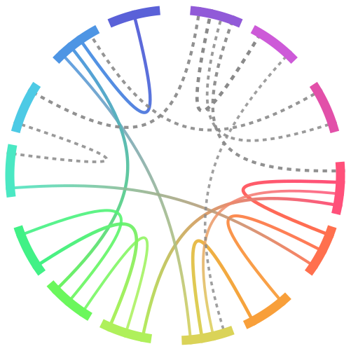
Network Atlas
Six brain states drawn as a chord ring — flow, rest, focused attention, threat, reward pursuit, meditation. Click a node, or a whole system, and the same regions light up on the scan beside it in the same colours.
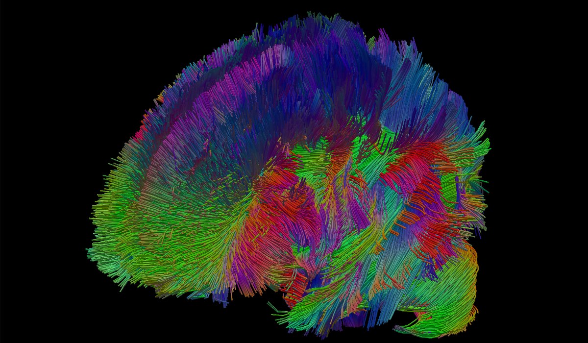
Tractography
The HCP1065 population-average tractogram: a million estimated fibre paths through the white matter, coloured by the direction each one runs and turned freely in 3D.
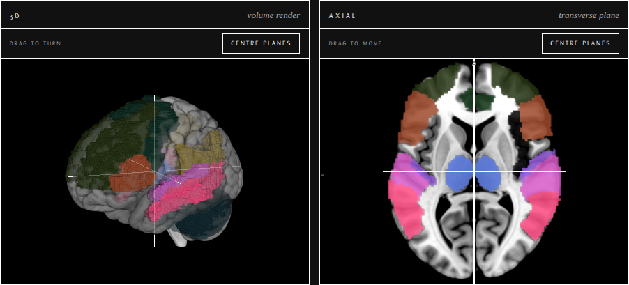
Visualizing Studies
Published work read through the brain map. Each study takes the regions its authors name, draws them on the scan where they actually sit, and sets out what the imaging found alongside what it does not settle — the reservations travel with the findings.
Now showing: Schizophrenic Hallucinations — ten regions of the auditory, language and control systems, switched between normal audition and the hallucinating state.
Microanatomy
The brain at the cell's scale. Original drawings built from code, beginning with a large multipolar neuron and the axons, astrocyte and synapses that meet it. Every structure is a click target — dendrites and spines, the cell body and what fills it, the hillock and initial segment, the axon under its sheath and the nodes between. Read what each one does, colour them all at once, or test yourself and find every structure from memory. Below the cells, the neurotransmitters, each drawn as a skeletal formula, and the hundred and fifty receptors, channels and transporters they act through, each with its kind of machine drawn and what binds it.
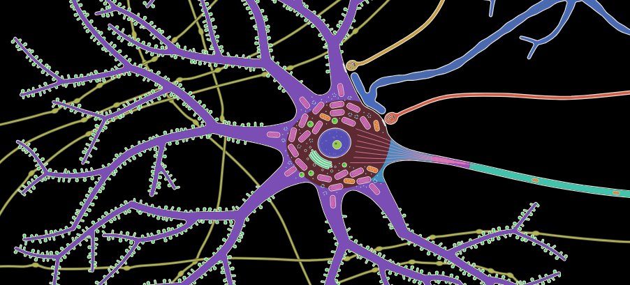
Molecular
The receptors themselves, in three dimensions. The five dopamine receptors and the twelve serotonin receptors that signal through G proteins, each built from experimental structures. Pick one and follow its neurotransmitter in as it docks and switches the receptor on, step by step, with the G protein docking after it. See where antipsychotics, Parkinson’s agonists, triptans, LSD and psilocin sit in the same pocket and whether they block the receptor or mimic its transmitter, then compare every subtype of a family side by side, pocket residue by pocket residue.
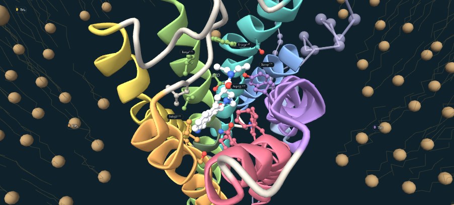
Neuroscience Practice
Adaptive multiple-choice practice built for the KCL MSc Neuroscience syllabus. It scores the session as you go, leans towards the topics you are getting wrong, and explains each answer — made to be worked through on a phone between lectures.
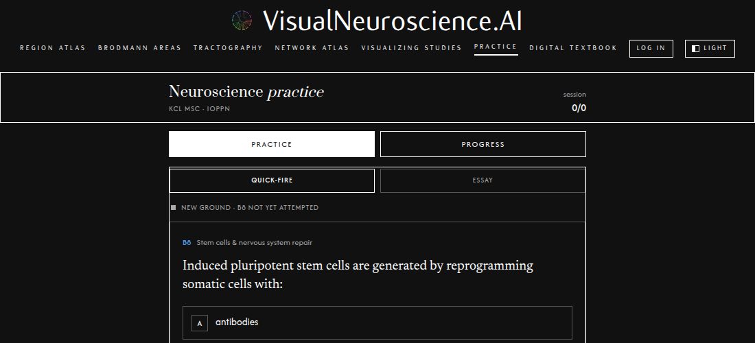
Digital Textbook
OpenStax’s Introduction to Behavioral Neuroscience, the whole book, set in this site’s own pages: nineteen chapters and eighty-five sections from the cells of the nervous system to attention and executive function, with every figure, note, key term and reference list. Chapters fold open to their sections and sections fold open to their text, so it reads on a phone as well as a desk. Free and openly licensed, as the original is.
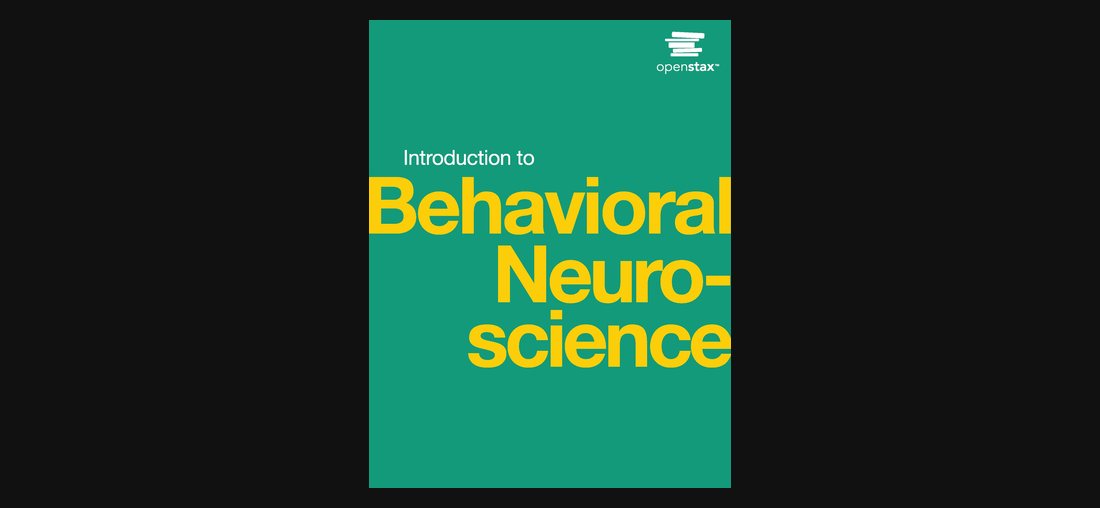
Everything here runs in the page: the volumes, the atlas and the tractogram are shipped as files and rendered on your own machine, so nothing you look at leaves it. Rendered with NiiVue (Rorden lab) · MNI152 template · AAL atlas · set in Flux, Newsreader & Prata · © 2026 VisualNeuroscience.AI About · Privacy & support.